Full Mouth Implant Rehabilitation for a 65-Year-Old with Decayed Teeth, Missing Teeth, and Uneven Bite
Doctor’s Name | Dr. Namratha Chandrahari |
Patient’s Name | Rabiya Bashri |
Chief Complaint | Missing teeth in the upper and lower jaw, decayed teeth, and improper occlusion on the right side, causing difficulty in chewing |
Diagnostic Method | CBCT (Cone Beam Computed Tomography) |
Clinic Name | Enamel Dental Clinic, Banjara Hills, Hyderabad |
Patient’s Age | 65 years |
Gender | Female |
Treatment | Full Mouth Rehabilitation — 5 Upper Jaw Implants + 2 Lower Left Implants, DMLS Fixed Teeth on the Upper Jaw, Tooth-Supported Zirconia Crowns and Prosthesis on the Lower Jaw |
Patient Background
Rabiya Bashri, a 65-year-old patient, came to Dr. Namratha Chandrahari, widely regarded as the best implantologist in Banjara Hills, with a mouth that had been failing her for years. Several teeth were missing across both the upper and lower jaw, several more were badly decayed, and her bite on the right side had shifted out of alignment. The combined effect was a mouth that could no longer chew properly, forcing her to favour the left side and avoid many foods altogether.
A single tooth, or even a few crowns, was never going to resolve what Rabiya was dealing with. This was a case of partial edentulism spread across both arches, layered on top of decay and an occlusion that had drifted over time. Following a detailed CBCT assessment, Dr. Namratha recommended a full-mouth rehabilitation plan that would replace what was missing, remove what was beyond saving, and rebuild a bite that worked evenly across both sides again.
Presenting Symptoms
The clinical presentation documented by Dr. Namratha Chandrahari captured the following findings across Rabiya’s case:
- Missing teeth in both arches. Rabiya had lost multiple teeth across the upper and lower jaw over the years, leaving functional gaps on both sides.
- Decayed teeth. Several of the remaining natural teeth showed significant decay, with some root structure no longer restorable with a filling or crown.
- Improper occlusion on the right side. The bite on the right side had shifted out of alignment, placing uneven load on the remaining teeth and the jaw joint.
- Difficulty in mastication. With missing and decayed teeth on both arches, Rabiya was struggling to chew food properly and had shifted most of her chewing to whichever side still offered some support.
- Partially edentulous arches. The combination of tooth loss in both jaws meant the case needed to be planned as a full arch problem rather than a series of isolated gaps.
Diagnostic Method
A thorough clinical examination was carried out to assess the extent of tooth loss, the condition of the decayed teeth, and the degree of occlusal misalignment on the right side. A CBCT (Cone Beam Computed Tomography) scan was taken of both the upper and lower jaw to build a complete three-dimensional picture of the available bone.
The scan measured bone height, width, and density at each proposed implant site across the upper arch and the lower left quadrant, and mapped the position of the sinus floor and surrounding anatomical structures. In a full-mouthcase involving implants across two arches, this level of imaging is what allows the number, position, and angulation of implants to be planned precisely before any surgical step is taken.
Diagnosis
Clinical examination together with CBCT imaging confirmed a diagnosis of partial edentulism affecting both the upper and lower jaws, with associated dental decay and improper occlusion on the right side. The findings indicated that a coordinated, implant-supported full-mouth rehabilitation rather than isolated single-tooth solutions would give Rabiya a stable, evenly distributed bite and a predictable long-term result.
Treatment Plan
A comprehensive full mouth rehabilitation plan was developed for both arches, incorporating the following clinical steps:
- Preoperative bone assessment and surgical mapping: CBCT data was used to plan implant positions across the upper arch and the lower left quadrant, identifying sites with sufficient bone volume for direct implant placement.
- Removal of non-restorable decayed teeth: Teeth and root structure that could not be saved were extracted as part of the preparatory phase, clearing the way for implant planning.
- Placement of 5 implants in the upper jaw: Five implants were placed across the upper arch at the planned positions, forming the foundation for a fixed, implant-supported restoration.
- Placement of 2 implants in the lower left jaw: Two additional implants were placed in the lower left quadrant to restore the missing teeth in that region.
- DMLS fixed prosthesis on the upper jaw: A Direct Metal Laser Sintering (DMLS) framework was planned for the upper arch, offering a precisely fitted, implant-supported fixed restoration as part of a broader full mouth dental implants treatment plan.
- Tooth-supported zirconia crowns and prosthesis on the lower jaw: The remaining healthy teeth in the lower jaw were used to support zirconia crowns and a connected prosthesis, following the same principle used in full mouth reconstruction cases, where implants, crowns, and natural teeth are brought together into one functioning bite.
- Healing period and monitoring: A structured healing period was built into the plan to allow osseointegration at every implant site before final restorative work proceeded.
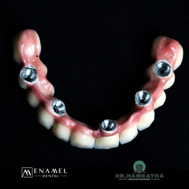
Laboratory-fabricated implant-supported prosthesis — screw access view
Surgical Procedure
The procedure was carried out under local anaesthesia with strict aseptic technique throughout. Non-restorable decayed teeth were extracted first, and each site was assessed against the CBCT plan before implant placement proceeded.
Five implants were placed across the upper arch, and two implants were placed in the lower left quadrant, with careful attention paid to spacing and angulation so that the eventual crowns and prosthesis would sit in proper alignment with the opposing arch. Primary stability was checked at each site before the healing phase began. Intraoral photographs taken during treatment confirmed accurate implant position across the upper arch before the fixed prosthesis was fitted.
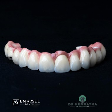
Finished zirconia crown and prosthesis — lower jaw
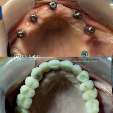
Upper jaw implants placed and final crowns — occlusal view
Post-Surgery Care and Guidelines
Detailed post-operative instructions were issued by Dr. Namratha Chandrahari to guide Rabiya through healing and protect the integrity of the newly placed implants:
- Oral hygiene protocol: Gentle brushing with a soft-bristled brush was advised from the second day after surgery, along with prescribed antiseptic mouth rinses for the first two weeks.
- Dietary guidelines: A soft diet was recommended through the healing period, with firmer foods reintroduced gradually once healing was confirmed at review.
- Medication compliance: Prescribed antibiotics were to be completed in full, with pain relief taken as needed to manage post-operative discomfort.
- Behavioural restrictions: Smoking and any habits that could interfere with healing were to be avoided while the implant sites integrated.
- Occlusal monitoring: Given the pre-existing improper occlusion on the right side, bite alignment was checked closely at each review visit as the fixed restorations were fitted.
- Long-term follow-up proFFtocol: Review appointments were planned to monitor healing, confirm integration at all seven implant sites, and check the fit of the final crowns and prosthesis once placed.
Outcomes & Results
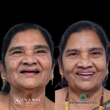
The treatment outcome for Rabiya was clinically excellent. The implants placed across the upper jaw and lower left quadrant showed satisfactory healing and stable integration at review, with a bite that was evenly distributed and no longer tilted toward the previously affected right side. The DMLS-supported fixed teeth on the upper jaw and the tooth-supported zirconia crowns and prosthesis on the lower jaw gave Rabiya a stable occlusion, restoring both function and appearance.
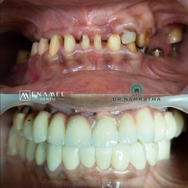
Before and after intraoral view — decayed and missing front teeth restored with a fixed full-arch prosthesis
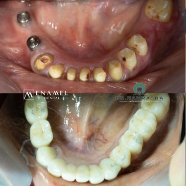
Before and after occlusal view of the lower jaw — decayed teeth and implant sites restored with zirconia crowns
The overall clinical assessment, conducted by Dr. Namratha Chandrahari, recorded an excellent prognosis, with satisfactory healing and stable occlusion achieved across both arches.
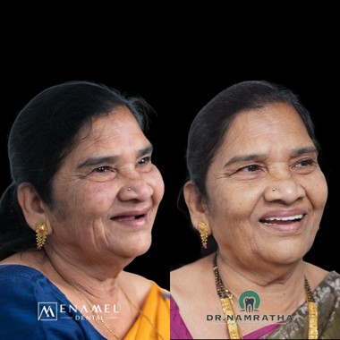
Before and after — profile view smile comparison
Long-Term Expectations
With consistent oral hygiene and the recommended review schedule, the outlook for Rabiya’s full mouth rehabilitation is excellent. The seven implants placed across the upper jaw and lower left quadrant function as stable, bone-anchored support for the fixed restorations, and the zirconia crowns on the lower jaw are expected to hold up well under normal biting load over the years ahead. Periodic professional cleaning around the implant-crown margins, along with regular check-ups with Dr. Namratha Chandrahari, will help catch any issues early and keep the stable occlusion performing well long-term.
Book Your Appointment
Dealing with a damaged tooth and old root stumps you’ve been putting off?